Two-photon laser-scanning fluorescence microscopy has become a well-established technique for high-resolution imaging from living tissue. However, standard microscopes are inflexible and the imaging depth is limited to a depth of 1 mm from the tissue surface. Several applications in the biomedical field, such as optical biopsy or brain imaging in freely behaving animals, require small, flexible instruments to overcome these limitations. A research team from the Max-Planck-Institut für Medizinische Forschung (Heidelberg, Germany) has developed a miniaturized two-photon microscope, which promises to enable flexible endoscopic imaging from deep within various tissues.1 The system was used for in vivo imaging of fluorescently labeled blood vessels in rat.
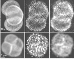
The probe of a compact two-photon microscope is an imaging fiber bundle. Pollen-grain images from a standard two-photon microscope (left) are compared with raw images from the fiber-bundle microscope (center) and images from the new microscope after smoothing with a Gaussian-blur filter (right).
Two-photon-fluorescence microscopy requires a way of delivering the excitation-laser light to the sample, scanning the focused light across the sample, and collecting the resulting fluorescence. The Heidelberg team, headed by Fritjof Helmchen, designed a system that uses a thin optical-image guide that contains several thousand fibers, allowing the coherent transmission of images back from the sample. A major advantage of such fiber bundles for miniaturization is that the scanning mechanism can be placed at the fiber entrance within the instrument; the alternative single-fiber approach requires a bulky scanner at the fiber exit at the sample end. With a fused coherent bundle, the input beam can be scanned across the fibers and fluorescence gathered through the whole bundle.
Prechirped pulses
The system uses 880-nm laser pulses with an initial pulse length of approximately 100 fs, from a modelocked Ti:sapphire laser (see schematic). A Pockels cell controls intensity. The pulses are negatively prechirped using a double pass through a pair of diffraction gratings to precompensate the positive group-velocity dispersion that occurs in the fiber-bundle cores. Despite this, pulse broadening is seen at higher pulse energies. The laser beam is then coupled to a custom laser-scanning microscope and focused onto the fiber-bundle entrance face. Raster scanning results in sequential coupling of the laser beam into individual fiber cores with a coupling efficiency of around 50%.
The fused coherent fiber bundle is 140 cm long and is composed of 30,000 individual cores, each with a core diameter of 2.4 μm and a numerical aperture of 0.35. The distance between two neighboring cores is 4 μm. The image area (the area containing fiber cores) has a diameter of 0.8 mm; the total outer diameter of the bundle, including its protective jacket, is 0.95 mm.
The output pattern of the fiber bundle is demagnified using a 1-mm-thick gradient-index (GRIN) lens attached to the fiber-bundle endface, resulting in a maximum field of view of about 300-µm diameter. Fluorescent light is collected back through the GRIN-lens objective and the fiber bundle, separated from the IR excitation light, and then detected by a photomultiplier tube.
The acquired images appear pixelated; a smoothed visualization was achieved by digitally blurring the images with a Gaussian filter. This effect was demonstrated with images of pollen grains (see photo).
An important aim was the ability to obtain images from intact living tissue. As an initial step in this direction, the system was applied to in vivo imaging in the brain of anesthetized rats. To enable imaging of blood vessels, the blood plasma was fluorescently labeled by tail-vein injection of Fluorescein or Rhodamine dye. The GRIN-lens objective (the tip of the fiber-bundle microscope) was positioned on the exposed brain surface and images of blood vessels of various sizes obtained (see photograph).
“Our system based on a coherent fiber bundle and a GRIN-lens objective is both small and light,” says Helmchen. “For the future, we expect that fluorescence excitation can be improved by optimizing the laser-pulse shape, and that the detected signal can be further increased by collecting fluorescence through a separate large-core detection fiber. The fiber-bundle microscope promises flexible endoscopic imaging from deep within various tissues with many applications in biomedical research.”
REFERENCE
1. W. Göbel et al., Optics Lett. 29(21) 2521 (Nov. 1, 2004).
