Custom precision rotary table enables fastest 3D x-ray tomographic images
Researchers at Helmholtz Zentrum Berlin (HZB) have developed a precision rotary table at the EDDI beamline at BESSY II and combined it with a fast CMOS camera, enabling them to document the formation of pores in grains of metal during foaming processes at 25 x-ray tomographic images per second (a world record).1
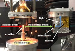
The quality of materials often depends on the manufacturing process. In casting and welding, for example, the rate at which melts solidify and the resulting microstructure of the alloy is important. With metallic foams as well, it depends on exactly how the foaming process takes place. To understand these processes fully requires fast sensing capability. Francisco Garcia-Moreno and his team have designed a turntable that rotates ultrastably about its axis at a constant rotational speed. The experiment requires extremely high precision—any tumbling around the rotation axis or even minimal deviations in the rotation speed would prevent reliable calculation of the 3D tomography.
Custom-built rotary table While commercially available rotary tables costing several hundred thousand euros allow up to 20 tomographic images per second, the Berlin physicists were able to develop a significantly cheaper solution that is even faster. "My two doctoral students at the Technische Universität Berlin produced the specimen holders themselves on the lathe," says Garcia-Moreno. Additional components were produced in the HZB workshop. In addition, Garcia-Moreno and his colleague Catalina Jimenez had already developed specialized optics for the fast scientific CMOS camera during the preliminary stages of this work that allow for simultaneous diffraction at multiple wavelengths (the source is a "white" x-ray source, having a continuous spectral band), making it possible to record approximately 2000 projections per second, from which a total of 25 3D tomographic images can be created.
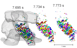
As a first example, the team investigated granules of aluminum alloys that become a metallic foam when heated. To do this, they mounted an infrared lamp above the metal granulate to heat the sample to about 650°C. A complete 3D tomographic image with spatial resolution of 2.5 µm (the pixel size) was generated every 40 ms. The nearly 400 tomographic 3D images allow detailed, time-resolved analysis of the process as it occurs."We wanted to develop a better understanding of how pores form in the grains—whether they also reach the granule surfaces and to what extent this process varies in different granules," says Garcia-Moreno.
Data is useful for industry
This is a question of practical relevance to industry, as granules of metallic compounds could fill complicated shapes better while foaming than foams manufactured from a block of metal. However, the molded part will only be able to withstand stress if the grains also bond closely with one another during foaming. With the ultrafast 3D tomography developed at BESSY II, this can now be observed very precisely.
Since the EDDI beamline must be dismantled for an upgrade from BESSY II to BESSY-VSR, Garcia-Moreno is in contact with other x-ray sources and plans to establish this method at those locations.
Source: http://www.helmholtz-berlin.de/pubbin/news_seite?nid=14865;sprache=en;typoid=3228
REFERENCE:
1. Francisco García-Moreno et al., Journal of Synchrotron Radiation (2018); https://doi.org/10.1107/S1600577518008949

John Wallace | Senior Technical Editor (1998-2022)
John Wallace was with Laser Focus World for nearly 25 years, retiring in late June 2022. He obtained a bachelor's degree in mechanical engineering and physics at Rutgers University and a master's in optical engineering at the University of Rochester. Before becoming an editor, John worked as an engineer at RCA, Exxon, Eastman Kodak, and GCA Corporation.

