Microscopy: Fourier-transform light scattering microscopy identifies individual bacteria

Researchers at the Korea Advanced Institute of Science and Technology (KAIST; Daejeon, South Korea) have developed a new way to exploit optical microscopy for identifying bacteria. Based on Fourier-transform light scattering (FTLS), the technique is not only label-free, but allows identification of a single bacterium.1 It is potentially important for the rapid identification of harmful pathogens that cause either human illness or the contamination of food.
The new approach involves bouncing laser light off individual bacteria under the microscope, using quantitative phase imaging (QPI) to create holographic images of them, and then using a software-based (machine learning) Fourier transform to analyze the images and identify them by comparing the Fourier-transform results to those from other known bacteria.
The setup includes a diffraction phase microscopy (DPM) component containing a common-path interferometer with off-axis spatial modulation that captures single-shot full-field images. The phase information extracted via QPI is essential to the technique.
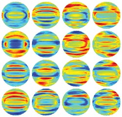
Information is extracted from the resulting 2D light-scattering spectra (with the "spectrum" being spatial frequency, not wavelength) from within the circular region of interest (ROI), which consists of 5525 pixels. A "feature extraction" procedure called principal-component analysis (PCA) then reduces the number of variables describing the ROI to 250 principal patterns, reducing the amount of computation needed while preserving 99.9% of the information.
A linear discriminant analysis (LDA) software model is then used to classify the species of the individual bacterium from which the FTLS and QPI information had been determined. The researchers examined four different bacterial species (Listeria monocytogenes, Escherichia coli, Lactobacillus casei, and Bacillus subtilis). The first three are all pathogens known to infect humans through the food chain or via hospital-acquired infections. The fourth is a harmless bacteria used in laboratory research, but of great interest because it is closely related to the deadly Bacillus anthracis, which causes anthrax.
Under a microscope, all four of these rod-like bacteria look nearly identical and would be virtually impossible to visually distinguish. The KAIST team, however, sorted them using the QPIU process with accuracy greater than 94%. As noted in the paper, "The overall cross-validation accuracy was 94.05%, with sensitivities of 95.52%, 95.65%, 88.46%, and 98.18% and specificities of 99.51%, 96.50%, 97.91%, and 98.13% for L. monocytogenes, E. coli, L. casei, and B. subtilis, respectively."
Conversion of existing microscopes
"We have also developed a compact portable device, so called quantitative phase imaging unit (QPIU),2 to convert an simple existing microscope to a holographic one, in order to measure light scattering patterns of individual bacteria, which can then be used to identify bacteria species for rural areas and developing countries," says physicist YongKeun Park. "Our team has plans to go to Tanzania next month for a field test."
The existing gold standard in the health-care industry for making a definitive diagnosis involves culturing the microbes from a patient's blood in the lab—a slow technique. Also routinely used today is a DNA-analysis technique called quantitative polymerase chain reaction (qPCR). This approach is faster, but can still take hours to produce a result (in addition, it requires expensive sample preparation that is often too expensive for routine use in rural or resource-poor settings around the world). The hours-long wait can be lethal in cases of sepsis or other fast-moving bacterial infections. As a result, many hospital-acquired infections are treated presumptively, before they are definitively identified, using broad-spectrum antibiotics. These powerful combinations of potent drugs are often effective, but using them routinely raises the risk of breeding deadly multidrug-resistant bacteria.
The KAIST researchers are now looking to extend their initial work to see if they can distinguish between several types of bacterial subgroups, allowing them to identify the most drug-resistant or virulent strains from the innocuous ones.
REFERENCES
1. Y. Jo et al., Opt. Express (2015); doi:10.1364/OE.23.015792.
2. K. Lee and Y. Park, Opt. Lett. (2014); doi:10.1364/OL.39.003630.

John Wallace | Senior Technical Editor (1998-2022)
John Wallace was with Laser Focus World for nearly 25 years, retiring in late June 2022. He obtained a bachelor's degree in mechanical engineering and physics at Rutgers University and a master's in optical engineering at the University of Rochester. Before becoming an editor, John worked as an engineer at RCA, Exxon, Eastman Kodak, and GCA Corporation.