BIOPHOTONIC IMAGING: Multispectral imaging targets pressure ulcers and deep-tissue injuries
Engineers at Georgia Institute of Technology (Georgia Tech; Atlanta, GA) and Georgia Tech Research Institute (GTRI; Atlanta, GA) have designed an enhanced multispectral imaging system for detecting bruising and erythema (skin reddening). The system consists of a laptop computer that imports images from a camera with a multispectral filter wheel. The camera’s filters measure narrow bands of visible light—about 10 nm in length. “The filters are highly sensitive to particular biological phenomena such as hemoglobin,” said Jayme Caspall, a GTRI research engineer.
Researchers shine a broad spectrum of light onto the skin and measure the light that reflects (see figure). They focus on the narrow bands because certain chemicals—like proteins in the blood near the skin surface—have peaks in absorption or reflectivity in certain wavelengths. For example, light is absorbed by hemoglobin, so if the scanner display goes dark, an accumulation of hemoglobin is present under the skin, indicating tissue injury.
Health-care professionals routinely check patients for early signs of erythema, or skin redness. But visual inspections sometimes fail to detect reddening of the skin and other indicators of tissue damage, especially in people with darkly pigmented skin. If undetected, these at-risk sites can develop pressure ulcers. Beyond ulcers looms a more serious risk for these patients—that of pressure-induced deep-tissue injury, which occurs below the skin and is often not diagnosed visually until it has reached a dangerous, advanced stage.
Aiming for low cost
Health-care practitioners may be able to reduce their patients’ risk of these complications by supplementing their visual inspections with a low-cost handheld imaging device that could detect early-stage pressure ulcers as well as the more serious deep-tissue injuries. Such a device is the ultimate goal of a Georgia Tech study now in field trials. The work is being funded by a grant from the National Institutes of Health (NIH; Bethesda, MD) in collaboration with the National Institute of Justice (NIJ; Washington, D.C.). A significant focus of the study is on detection of bruises and erythema in people with darkly pigmented skin.
“There’s a huge opportunity to intervene if we can see pressure ulcers at a very early stage,” said lead researcher Stephen Sprigle, director of the Georgia Tech College of Architecture’s Center for Assistive Technology and Environmental Access. “Detecting them then drives the treatment. If you take the visual indicator away, it adversely impacts care, and for folks with darkly pigmented skin, that’s a problem.” In addition to the human costs of pressure ulcers, there are monetary burdens. The cost to heal a pressure ulcer is estimated to reach $40,000 for certain ulcers, Sprigle notes. In the United States alone, the costs associated with healing ulcers and worker productivity losses exceed $2 billion a year.
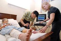
Clinical field studies began in January 2006 and will continue through the summer at a nursing home in Atlanta and at the Shepherd Center, an Atlanta-based catastrophic-care hospital. Because of immobility, patients at Shepherd and residents in the nursing home are at risk for pressure ulcers. Researchers hope to get data over weeks or months as healing occurs on about 25 to 30 patients, particularly those with dark skin.
Clinical data collection helps drive the researchers’ modeling and algorithm development. Though these studies don’t reveal the etiology and the time base on bruises, they will provide enough background information to do controlled animal studies later, Sprigle says.
The current phase of the project was funded through last month, and Sprigle plans to apply to NIJ and NIH for continuation funding. Ultimately, the researchers plan to develop a prototype software program and/or hardware device. The researchers estimate the ultimate product—a handheld imaging device—will cost no more than $5,000 per unit. They want to license the technology to an optics and/or acoustics firm.
“There’s a need for this technology in every nursing unit in every hospital, group home, nursing home and rehabilitation facility,” Sprigle said. “Also, if we can detect bruising caused by abuse, it will be useful to social service agencies.”
About the Author
Hassaun A. Jones-Bey
Senior Editor and Freelance Writer
Hassaun A. Jones-Bey was a senior editor and then freelance writer for Laser Focus World.