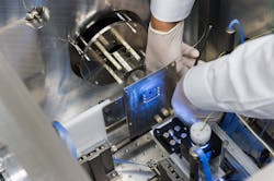
Cell Imaging/Cell Analysis: EUV-enabled spectrometry images cells in 3D at nanoscale
Researchers at Colorado State University (Fort Collins, CO) have developed a spectral imaging instrument that maps cellular composition in 3D at the nanoscale.1 The system allows study of cells at approximately 100% greater detail than previously possible, enabling observation of cell response to drugs.
The researchers explain that earlier laser-based mass-spectral imaging could identify the chemical composition of a cell and could map its surface in 2D at the microscale, but could not chart cellular anatomy in nanoscale or in 3D.
The instrument features a laser able to produce a hot, dense plasma stream that acts as a gain medium for generating extreme ultraviolet (EUV) pulses. When properly focused, the laser drills a tiny hole into a cell sample, enabling ions to evaporate from the cell surface. These ions can be separated and identified to determine chemical composition, and used to chart the anatomy of a cell.
1. I. Kuznetsov, NatureCommun., 6, 6944, doi:10.1038/ncomms7944 (2015).

Barbara Gefvert | Editor-in-Chief, BioOptics World (2008-2020)
Barbara G. Gefvert has been a science and technology editor and writer since 1987, and served as editor in chief on multiple publications, including Sensors magazine for nearly a decade.
