Tissue Imaging: Got nerve? Collimated polarized light can tell
Surgeons like to know the location of nerve tissue during operations, but too often, they don't. It's so hard to differentiate nerves—especially the smaller ones—from other tissue, as eyeballing by the surgeon has a success rate of only about 77%. And the methods thus far developed to make nerves more visible have major limitations, including need for fluorescent dye or lag time in delivering imagery.
But polarized light is showing the way to real-time, noninvasive visualization of nerve tissue. It promises to help surgeons quickly locate nerves needing repair and, perhaps more importantly, to avoid severing them by accident, leaving the patient with either chronic pain or reduced or lost function or sensation.
Hobbyist cousins' collaborative
The application is the brainchild of two then-amateur scientists who happen to be related. "What started out as a crazy hobby project with my cousin Patrick is now a new optical imaging technique that is capable of recognizing nerves in operations in real time, thus preventing nerve injury in patients," said Kenneth Chin. Chin made that statement in accepting the 2017 Amsterdam Science & Innovation High Potential Award, which recognized his entrepreneurship (with a €5,000 prize) based on the cousins' scientific work. The High Potential Award is one of three presented for 2017 by IXA, Innovation Exchange Amsterdam; the program honors innovative ideas based on scientific research with social and/or commercial application.
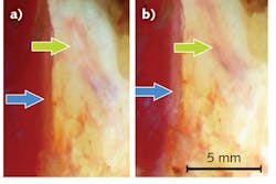
When Kenneth and Patrick tried using polarized light to illuminate tissue, they found that the internal structure of nerves reflected the light in a unique way: It relates to the nerve fiber's orientation compared to that of the light's polarization. And, they discovered that by rotating the light's polarization, they could get the reflection to appear to switch on and off, making the nerves stand out from other tissue. Further, they learned that use of collimated light (in which light waves are parallel) maximizes the reflection's strength. They call their approach collimated polarized light imaging (CPLi).
Prototype development and application
Following their discovery, Kenneth joined a research group led by Thomas van Gulik, a surgeon at the Academic Medical Center, University of Amsterdam, Netherlands. He brought to the university a working prototype, which the group has since developed into a practical system, using a long working distance for intraoperative use. "We adapted the optics used for CPLi so that they could be incorporated in a surgical microscope, which can be placed above the surgical area," Chin said. "The resulting system can be used in a wide range of surgical fields where superficial nerves need to be identified."
After testing the technique on animal tissue, the researchers used it to examine 13 tissue sites from the hand of a human cadaver. Although nerve injury is a risk for many types of surgery, hand and wrist procedures are more prone to peril because the nerve networks are so dense in these areas. In their study, one surgeon looked for nerve tissue by eye under typical surgical illumination, while another used CPLi for an independent assessment. The researchers then used histological evaluation to verify the presence of nerve tissue at each site. The surgeon using visual inspection correctly identified nerve tissue in 10 of the 13 cases, but the one using CPLi correctly identified nerve tissue in all cases.
The researchers subsequently used CPLi to successfully identify nerve tissue during a wrist procedure in a living patient. They plan additional intraoperative tests to better understand how the optical reflection of nerves might vary among patients and under differing surgical conditions. With further development, they hope for production of a clinically approved CPLi system.
REFERENCE
1. K. W. T. K. Chin et al., Biomed. Opt. Express, 8, 9, 4122–4134 (2017).

Barbara Gefvert | Editor-in-Chief, BioOptics World (2008-2020)
Barbara G. Gefvert has been a science and technology editor and writer since 1987, and served as editor in chief on multiple publications, including Sensors magazine for nearly a decade.
