Government funds aid optical imaging
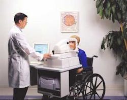
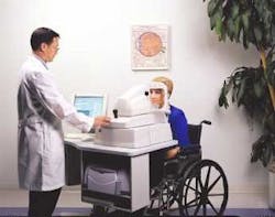
There are changes afoot in the hallways and boardrooms of the National Institutes of Health (NIH; Bethesda, MD), and they have the optical-imaging research community applauding. The operative word is "funding," and the U.S. government is putting its money where its mouth is and investing in new technologies and medical devices that might actually make it into the hands of physicians and improve the standard of medical care (see figure).
The ability of optical technologies to offer earlier, less invasive, more targeted disease detection has been well researched and documented; however, taking these same technologies from "benchtop to bedside" has been anything but easy. According to James Fujimoto, professor of engineering at Massachusetts Institute of Technology (MIT; Cambridge, MA) and a pioneer in the development of optical coherence tomography (OCT), medical instrumentation is challenging to develop and take to market because there are so many hurdles along the way, including demonstration studies, prototype development, feasibility studies, clinical trials, regulatory reviews, sales to early adopters, demonstration of clinical efficacy, reimbursement issues, and adoption by the clinical community at large.1 The result is often a very long and costly process that, despite an R&D team's initial enthusiasm, can easily lose steam—and funding—after a few years.
"In the past, SBIRs (small business innovation research grants) would often go through the feasibility and prototype development phase but no further," said Houston Baker, program director in the cancer-imaging program at the NIH's National Cancer Institute (Bethesda, MD). "Especially in medicine, once you have a prototype there is a lot still to be done to put it in the hands of a technician working with a physician who will use it on a patient. This is the whole concept of benchtop to clinic."
Baker and colleagues at NCI took this observation to heart and began designing a funding initiative that would help researchers and commercial groups share their expertise and move their inventions further along the product-development pipeline. The result was the Network for Translational Research for Optical Imaging (NTROI), a five-year NIH program that supports three to four multidisciplinary research teams focused on the development, validation, and translation of prototypes to clinically ready optical-imaging instruments and techniques for early cancer detection.
One of the groups participating in the NTROI is the Beckman Laser Institute (Irvine, CA). Using a $7 million NTROI grant, lead investigator Bruce Tromberg and his colleagues at Beckman are working with researchers from the University of California–San Francisco, the University of Pennsylvania, Harvard University/Massachusetts General Hospital, Dartmouth University, University of Illinois at Urbana-Champaign, and Siemens Corporate Research to speed development of diffuse optical imaging for cancer detection and diagnosis in the breast. The researchers hope to use this information to develop laser-based imaging devices that complement conventional modalities such as mammography.
"There are many opportunities for translational research in biomedical optics to take this technology from the benchtop to the bedside," Tromberg said. "People in this field are now capitalizing on the insights gained in basic studies of tissue and light interaction by engineering systems that can acquire disease-specific information and validate the technology in the clinical setting."
Other NTROI-funded projects include a collaboration between Boston University (lead investigator is Irving Bigio), University College of London, University of Pittsburgh, Optimum Technologies (Southbridge, MA), Pharmacyclics (Sunnyvale, CA), and DiametRx (Natick, MA) to develop an in-vivo contact probe for elastic-scattering spectroscopy to study cancers of the GI tract; and a group comprising researchers from the University of Pennsylvania (lead investigator is Wafik El-Deiry), Children's Hospital of Philadelphia, Fox Chase Cancer Center (Philadelphia, PA), LightForm (Hillsborough, NJ), 3-Dimensional Pharmaceuticals (Yardley, PA), Morphotech (Irvine, CA), Cell Pathways (Horsham, PA), Cephalon (West Chester, PA), 5Star Medical (New Bedford, MA), Versilant Nanotechnologies (Philadelphia, PA), and NIM to develop bioluminescent probes and molecular markers for early detection of colon cancer.
The NCI is also funding several biomedical optics projects outside of the NTROI. The George R. Harrison Spectroscopy Laboratory at MIT won a $7.2 million Bioengineering Research Partnership grant from the NCI last year to further develop and implement spectroscopic techniques for imaging and diagnosing dysplasia in the cervix and mouth, while researchers from University of California–Irvine received $2.9 million to develop a microelectromechanical system-enhanced OCT microscope for 3-D endoscopic imaging of respiratory, gastrointestinal, and urogenital tracts. Other government agencies jumping on the bandwagon include the National Institute of Biomedical Imaging and Bioengineering and the National Science Foundation; the latter recently gave the University of Texas at Austin $3.8 million to establish a graduate program in cellular and molecular imaging for diagnostic and therapeutic applications.
All of this government activity has stimulated parallel activity among commercial providers of diagnostic imaging equipment. Philips (Eindhoven, The Netherlands), GE Medical (Milwaukee, WI), and Siemens (Erlangen, Germany) are all investing in what they consider the next generation of diagnostic systems, helping to validate for the first time the role of optical technologies in medical imaging. Philips and GE are both partnering with Advanced Research Technologies (St. Laurent, Quebec) on optical molecular imaging, a major provider of contrast agents for molecular imaging, while Siemens is pursuing optical molecular imaging for disease detection and working with some of the NTROI-funded groups on cancer imaging. GE also purchased Amersham (Buckinghamshire, England).
Outside the imaging community, even chipmaker Intel is investing in biomedical optics. In a joint project with the Fred Hutchinson Cancer Research Center (Seattle, WA), Intel researchers are leveraging technology originally developed for microchip defect detection to build the Intel Raman Bioanalyzer System; the goal is to be able to identify various markers in blood samples for early cancer detection.
REFERENCES
- J. G. Fujimoto, Nature Biotechnology 21(11) (November 2003).

