BIOPHOTONICS: Quasi-CW ultrasound adds mechanical information to optical tomography
By introducing a quasi-continuous-wave (quasi-CW) ultrasound modulation scheme into an ultrasound optical-tomography (UOT) scanner, researchers at Texas A&M University (College Station, TX) have enabled collection of mechanical information from a tissue sample in addition to the optical information normally supplied by UOT.
Developed over a period of years through the cumulative contributions of numerous research groups, UOT extends the tissue penetration depth of optical tomography from millimeters to practically useful distances on the order of centimeters.
After traveling about 200 µm in dense optical tissue, photons tend to lose the memory of their initial direction, phase, and polarization due to multiple scattering events. Interaction with an ultrasound signal, however, can modulate the optical path by varying both the refractive index of the transmission medium and the periodic motion of the physical particles that scatter passing light, which leads to an optical-frequency shift-or “photon tagging”-of light that has passed through the ultrasound signal. Acousto-optic imaging is then enabled by measuring the interference pattern between photons from the same coherent source that have and have not interacted with the ultrasound signal.
Noise introduced into the system by factors such as light speckle, as well as Brownian and other motions of living tissues, tends to limit the potential spatial as well as temporal resolution obtainable from the relatively small signals obtained through UOT. This problem was addressed about three years ago by the introduction of a photorefractive crystal (PRC) to compensate for the noise effects using self-adaptive wavefront holography.1
The Texas A&M researchers have taken this work a step further by applying the ultrasound modulation in the form of a 1-ms-long focused burst. In addition to improving the signal-to-noise ratio of the PRC-based UOT system, this quasi-CW ultrasound modulation also enabled detection of the acoustic-radiation force, which provides mechanical information about the sample.2
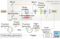
The researchers split 532 nm laser light into two paths, one for probing the sample in tandem with the ultrasound signal and the other for a reference signal to pump two wave mixing in the bismuth silicon oxide PRC (see figure). An alternating electric field (1 kHz, 4 kV) enhanced gain in the PRC, and pump-light intensity was maintained at approximately 10 mW/cm2. Measured crystal response time was on the order of 100 ms, and logarithmic improvement in UOT signal strength was observed with increases in ultrasound burst length. The ultrasound source was a focusing ultrasonic transducer with a central frequency of 1 MHz, focal length of 37.5 mm, focal-zone length of 23 mm, and focal-spot diameter of 2.2 mm. The peak acoustic pressure at the focus was 1.5 MPa and the duty cycle was 100 Hz.
The researchers demonstrated UOT imaging based on optical and mechanical properties by imaging segments of a nude mouse tail in a gelatin sample. The UOT image acquired 0.1 ms after the onset of the ultrasound provided optical absorption contrast. Mechanical characteristics of the sample, such as the acoustic impedance mismatch, which occur on a millisecond time scale, were obtained from time-evolution curves of the UOT signal. Time gating the optical signal also allowed decoupling of optical and mechanical information.
REFERENCES
1. F. Ramaz et al., Optics Express 12(22) 5469 (Nov. 1, 2004.
2. X. Xu et al., Optics Letters 32(6) 656 (March 15, 2007).
About the Author
Hassaun A. Jones-Bey
Senior Editor and Freelance Writer
Hassaun A. Jones-Bey was a senior editor and then freelance writer for Laser Focus World.
