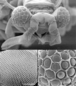
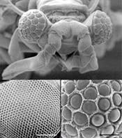
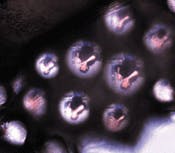
A group of scientists at Cornell University (Ithaca, NY), led by neurobiology professor Ron Hoy, has discovered an insect eye unlike any other. Belonging to Xenos peckii, a tiny parasite of wasps (top), the eye's design combines the omnidirectional properties of a compound eye with the higher resolution of single-lens eyes. An ordinary compound insect eye such as that of a fruit fly contains hundreds of individual elements (center left), each of which detects a single point in the field of view; thus, its resolution is never greater than the number of elements it contains. In contrast, the eye of Xenos peckii contains 50 or so refractive lenses, each capable of imaging a field of many points (center right). Such an eye is potentially limited in resolution only by its much higher number of retinal receptors. (In reality, lens performance comes into play, too.)
Because the eye of Xenos peckii is dome-shaped, the fields captured by the eyelets have little or no overlapa scheme ideal for omnidirectional imaging. However, because of the image reversal caused by the eyelets, the overall image created by the eye consists of many smaller inverted image sections that do not mesh together to form a coherent overall image. In a human-engineered optical system, this sort of problem would most likely be solved through specialized software or hardware. In the Xenos peckii eye, Nature decided on a hardware fix: behind each eyelet, the bundle of neurons carrying the optical signals undergoes a 180° twist before it joins with the other bundles at a neuronal receiving structure called the lamina. The resulting image information supplied to the lamina is thus free of image-reversed segments.
A typical eyelet is 65 µm in diameter, has a focal length of 45 µm, and captures an angular field of 33°. By removing a Xenos peckii eyelet array and flattening it on a microscope slide, the Cornell researchers were able to view its imaging properties (bottom), although flattening the array caused all the eyelets to image the same field, rather than the separate field segments that would be seen by the insect.
About the Author
John Wallace
Senior Technical Editor (1998-2022)
John Wallace was with Laser Focus World for nearly 25 years, retiring in late June 2022. He obtained a bachelor's degree in mechanical engineering and physics at Rutgers University and a master's in optical engineering at the University of Rochester. Before becoming an editor, John worked as an engineer at RCA, Exxon, Eastman Kodak, and GCA Corporation.


