Webcam-based hospital-grade laser-speckle blood-flow imager is low in cost
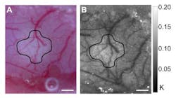
| A speckle contrast image (right) of a stroke-affected section of mouse brain averaged over 10 frames, with an image depicting the vascular anatomy (left) for comparison. (Credit: Andrew Dunn, University of Texas - Austin) |
Austin, Texas--Blood flow is routinely measured in the clinic and in the laboratory, and laser-speckle contrast imaging (LSCI) is one way of measuring this; however, this technique requires professional-grade imaging equipment, which limits its use. Now, using $90 worth of off-the-shelf commercial parts that include a webcam and a laser pointer, researchers at the University of Texas at Austin (UT-Austin) have duplicated the performance of expensive, scientific-grade LSCI instruments at a fraction of the cost.
The work is the first to show that it is possible to make a reliable blood-flow-imaging system solely with inexpensive parts, the authors say. The researchers describe their development in the latest issue of the Optical Society's (OSA's) journal Biomedical Optics Express.1
Measuring blood flow changes using LSCI requires laser light to illuminate the tissue, a camera to record the image, and focusing optics to direct the scattered light to the camera -- all normally at a price that can reach $5000.
Light source is ordinary laser pointer
In the UT-Austin approach, a $5 laser pointer is used to illuminate the patch of tissue to be imaged, and light is reflected off the tissue and focused by a pair of generic 40 mm camera lenses onto the sensor of a $35 webcam. The team used their setup to image changes in blood flow in a mouse model and found that they were able to identify areas of high flow versus low flow.
At just 5.6 cm in length and weighing only 25 g, the system is compact and lightweight, which would make it easier to transport for imaging applications outside of the lab, including clinics in areas with limited access to medical care, says biomedical engineer Andrew Dunn, an associate professor at UT-Austin and an author of the study. Currently, the new system's field of view, which is just a few square millimeters across, and resolution are limited by the size of the webcam sensor, but future versions could have a more flexible layout.
"The lens configuration could be easily adjusted to visualize even smaller structures by magnifying or to visualize larger structures by increasing the field of view, depending on what kind of flow the user is interested in visualizing," says Dunn.
REFERENCE:
1. L. Richards et al., Biomedical Optics Express, Vol. 4, Issue 10, pp. 2269-2283 (2013).
