OCT tracks growth and shrinkage of cone-cell appendages in living human eye
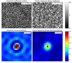
Bloomington, IN--Information hidden within optical coherence tomography (OCT) data is allowing researchers at Indiana University in Bloomington to track size changes of the "outer segments" of the eye's color-sensing cone cells.1 The so-called outer segment, made up of a series of stacked discs, each about 30 nm thick, is the light-sensing portion of the cone cell; it goes through daily changes in length (visible as changes in optical path length) that could help medical researchers identify potential retinal problems.
To make an OCT scan of the retina, a beam of light is split: one portion is scattered off the retina while the other is the reference beam. The beams are interfered, and the resulting phase information used to procure a precise measurement of a sample’s position. For this experiment, the researchers used an adaptive-optics (AO) OCT setup. But since in this case their samples were attached to live people, the researchers had to adapt the to counteract any movements that the subjects’ eyes might insert into the data.
Instead of measuring the phase of a single interference pattern, the researchers measured phase differences between patterns originating from two reference points within the retinal cells: the top and bottom of the outer segment. The team used this hidden phase information to measure microscopic changes in hundreds of cones, over a matter of hours, in two test subjects with normal vision. Researchers found they could resolve the changes in length down to about 45 nm, which is just slightly longer than the thickness of a single one of the stacked discs that make up the outer segment. The work shows that the outer segments of the cone cells grow at a rate of about 150 nm per hour.
REFERENCE:
1. Ravi S. Jonnal et al., Biomedical Optics Express, Vol. 3, Issue 1, pp. 104-124 (2012)
http://dx.doi.org/10.1364/BOE.3.000104

John Wallace | Senior Technical Editor (1998-2022)
John Wallace was with Laser Focus World for nearly 25 years, retiring in late June 2022. He obtained a bachelor's degree in mechanical engineering and physics at Rutgers University and a master's in optical engineering at the University of Rochester. Before becoming an editor, John worked as an engineer at RCA, Exxon, Eastman Kodak, and GCA Corporation.
