Confocal light-sheet microscopy is sharper way to view the brain's neural network
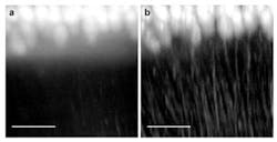
| Purkinje cells from a mouse cerebellum are imaged with light-sheet microscopy (a) and with confocal light-sheet microscopy (b). The scale bar at the bottom is 100 micrometers across. (Image: Optics Express/European Laboratory for Non-Linear Spectroscopy, University of Florence, Italy) |
Florence, Italy--A group of Italian researchers has combined light-sheet microscopy (LSM) with confocal microscopy to create a new form of microscopy that has the 3D imaging ability of LSM and the elimination of background scatter of confocal microscopy.1 Called confocal light-sheet microscopy (CLSM), the technique produces images 100% sharper than those acquired with conventional LSM, say the researchers.
The group includes scientists from the University of Florence, the University Campus Bio-Medico of Rome, the University of Cassino, the National Research Council, and the International Center of Computational Neurophotonics.
In LSM, a biological sample is illuminated with a thin sheet of light provided by a laser beam narrowed to just a few microns; the illumination comes from the side rather than from above or below as with traditional light sources. Optics and a digital camera capture the resulting fluorescence from the illuminated sample; many 2D slices of the sample are combined to create a 3D map of the sample. Adding confocal microscopy allows the system to capture light (point by point) from the fluorescing plane while rejecting scatter in tissue above and below the plane.
No postprocessing required
The combined system "filtered the scattered photons that were emitted and recovered the normally lost image contrast in real time without the need for multiple acquisitions or any postprocessing of the acquired data," says Francesco Pavone, the team leader. "This allows us to obtain sharp image slices that can be easily reconstructed into a macroscopic view of whole organs or entire organisms with a resolution of a few microns and faster acquisition time."
One region that scientists have tried to survey in mice with LSM is the neural pathway, the network of neurons that underlie the functioning of the brain. While the LSM method yields high-resolution views of tissue excised from mouse brains and fixed in position, whole-brain samples scatter the emitted light and create background fluorescence that reduces contrast and blurs the perceived image.
The researchers validated the background rejection capabilities of CLSM by showing that it results in significantly sharper images than conventional LSM microscopy and than a variation of LSM that requires redundant slices and postprocessing to remove scattered light. CLSM proved superior to the other two methods when used to view three samples from mouse brains: the hippocampus, the cerebellum, and the whole brain.
The 3D reconstruction ability of CLSM also was put to the test when the researchers used it to map the micron-scale neuroanatomy of mouse Purkinje cells (large neurons found in the cerebellum) and trace an entire brain's neuronal projections (the finger-like ends of a neuron by which neurochemical signals are received from other nerves).
Ludovico Silvestri, another member of the research team, says the group is using CLSM to image mouse models of several diseases, including autism and ischemic stroke. "We hope that the whole-brain tomographies we can obtain will one day provide insights into the mechanisms of these and other brain disorders," he says.
REFERENCE:
1. Silvestri et al., Optics Express, Vol. 20, Issue 18, pp. 20582-20598 (2012).
