HIGH-RESOLUTION BIOIMAGING/NANOTECHNOLOGY: Nanodiamonds enable magnetic resonance of individual cells, other bioimaging possibilities
Spanish and Australian researchers have collaborated on a new imaging technique similar to magnetic resonance imaging (MRI), but offering resolution and sensitivity sufficient to scan individual cells.1 A paper describing their work explains how artificial atoms—diamond nanoparticles doped with nitrogen impurity—can probe very weak magnetic fields such as those generated in some biological molecules. To trap and manipulate these artificial atoms, the researchers use laser light. The laser works like tweezers, leading the atoms above the surface of the object to study and extract information from its tiny magnetic fields. This effort represents the first time nanodiamonds have been optically trapped and manipulated in three dimensions.
Conventional MRI has a diagnostic resolution on a millimetric scale. The new technique provides nanometer resolution (nearly one million times greater), making it possible to measure magnetic fields created by proteins, for instance. It could revolutionize the field of medical imaging, allowing for substantially higher sensitivity in clinical analysis, an improved capacity for early detection of diseases, and thus a higher probability for successful treatment. The approach "will offer new sources of information and allow us to better understand the intracellular processes, enabling noninvasive diagnosis," explains Michael Geiselmann, who conducted the experiment. Geiselmann is with the Institute of Photonic Sciences (ICFO)—an associate institute of the Universitat Politècnica de Catalunya BarcelonaTech (UPC). The Spanish National Research Council (CSIC) and Macquarie University (Sydney, Australia) also contributed to the project.
Individual atoms are highly sensitive to their environment, with a great ability to detect nearby electromagnetic fields. But atoms are so small and volatile that in order to be manipulated, they must be cooled to temperatures near the absolute zero (about -273°C). This complex process requires an environment so restrictive that it makes individual atoms unviable for medical applications.
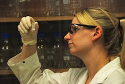
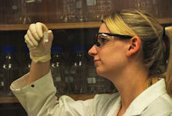
The team's artificial atoms are formed by a nitrogen impurity captured within a small diamond crystal. "This impurity has the same sensitivity as an individual atom but is very stable at room temperature due to its encapsulation," said Professor Romain Quidant of ICFO, whose team proposed the technique. According to Jana Say, a Macquarie PhD student, diamonds are more suitable than other nanoparticles for many biological applications because they are made from carbon, and thus are non-toxic. "We are working with nanodiamonds to process them so that they are stable enough to be used as a probe for single-molecule interactions," said Say.
Say's processing technique consistently generates stable products. "The real challenge is reliably producing the same sample," she noted. Under the supervision of Professor Louise Brown of the Department of Chemistry and Biomolecular Sciences, Say plans to continue to develop these diamonds and collaborate with other researchers to explore their full potential. Brown said that a recent project won a Macquarie research excellence award for demonstrating that Say's nanodiamonds can be isolated and made to emit light. "We continue to make real breakthroughs in this area and are contributing to the long-term goals in ultrasensitive imaging and sensing technologies," Brown added.
1. M. Geiselmann et al., Nat. Nanotechnol., doi:10.1038/nnano.2012.259 (2013).

