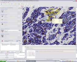
Enabling quantitative cell analysis in cancer research, clinical research, and pathology, TissuemorphDP image analysis software from Olympus (Center Valley, PA) detects and classifies cell nuclei, cytoplasm, or membrane. The software tool, designed by Visiopharm, allows users to control detection of cell structures via manual control of sensitivity and size. Able to customize thresholds for positivity for cell nuclei, research findings are stored electronically as user-defined protocols.
More Products
-----
PRESS RELEASE
Olympus Offers TissuemorphDP™ Cell Analysis Software Module from Visiopharm
CENTER VALLEY, Pa., January 3, 2012 – Scientists in the field of cancer research, clinical research and pathology will benefit from a dedicated software module available from Olympus that facilitates key aspects of working with quantitative cell analysis. TissuemorphDP image analysis software is specifically designed for detection and/or classification of cell nuclei, cytoplasm or membrane. Thanks to the tool’s easy-to-use preset protocols, many scientists will be able to load their samples and run complex analyses with the touch of just a few keys.
The software tool, designed by Visiopharm, allows users to control detection of cell structures through simple manual control of sensitivity (positive/negative nuclei and membrane) and/or size (positive/negative nuclei and cytoplasm). Users can customize thresholds for positivity for cell nuclei. Research findings and are stored electronically as user-defined protocols that can be loaded and executed easily by any authorized operator.
For more information on the TissuemorphDP cell analysis software tool, contact Olympus America Inc., 3500 Corporate Park Drive, Center Valley, PA 18034; phone Pia Rinta-Panttila at 1-484-896-5292, email [email protected] or visit www.olympusamerica.com/tissuemorph.
About Olympus America Inc., Scientific Equipment Group
Olympus America Scientific Equipment Group provides innovative microscope imaging solutions for researchers, doctors, clinicians and educators. Olympus microscope systems offer unsurpassed optics, superior construction and system versatility to meet the ever-changing needs of microscopists, paving the way for future advances in life science.
About Olympus
Olympus is a precision technology leader, designing and delivering innovative solutions in its core business areas: Medical and Surgical Products, Life Science Imaging Systems, Industrial Measurement and Imaging Instruments and Cameras and Audio Products. Olympus works collaboratively with its customers and affiliates worldwide to leverage R&D investment in precision technology and manufacturing processes across diverse business lines. These include:
Gastrointestinal endoscopes, accessories, and minimally invasive surgical products;
Advanced research, clinical and educational microscopes and research and educational digital imaging systems;
Industrial research, engineering, test, inspection and measuring instruments; and
Digital cameras and voice recorders.
Olympus serves the healthcare field with integrated product solutions and financial, educational and consulting services that help customers to efficiently, reliably and more easily achieve exceptional results. Olympus develops breakthrough technologies with revolutionary product design and functionality for the consumer and professional photography markets, and also is the leader in gastrointestinal endoscopy and clinical and educational microscopes. For more information, visit www.olympusamerica.com.
-----
Follow us on Twitter
Subscribe now to Laser Focus World magazine; it's free!
