Laser Doppler-driven camera shows blood circulating
Researchers at the École Polytechnique Fédérale de Lausanne (EPFL) and EFPL startup Aïmago (both in Lausanne, Switzerland) have developed a medical diagnostic imaging device that shows how blood is circulating in the skin in real time, enabling burn specialists to look at the quality of dermal blood circulation to judge a burn’s extent and severity. Other areas of medicine such as plastic surgery, wound healing, diabetes, rheumatology, and neurosurgery could also benefit from the device.
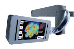
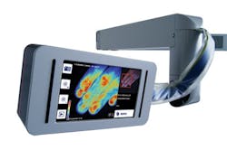
The device, invented in EPFL’s Biomedical Optics Laboratory (LOB) and named the EasyLDI, is based on laser Doppler imaging (LDI). First, an 808 nm single-mode diode laser beam at 150 mW is reflected by red blood cells and static tissue. Then, taking advantage of the Doppler effect—the frequency lag between the emitted and reflected light beam—a custom camera that incorporates a high-speed CMOS image sensor and can obtain 20,000 images per second transforms the microcirculation in the tissue into color variations on the interface.
When the device is held above the zone in question, microcirculation is displayed like a topographical map on the screen. The color variations it yields reveal differences in microcirculation intensity over a volume of skin about 50 cm2 in area and 1–2 mm deep.
Covered with a sterile sleeve, the device can also be used in the operating room. And when asked whether or not the device could become portable, Michael Friedrich, Aïmago president and CEO, told BioOptics World that this could be possible in a couple of years.
More BioOptics World Current Issue Articles
More BioOptics World Archives Issue Articles

