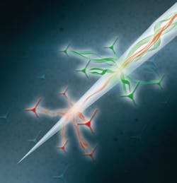
Tapered optical probe helps detect light in the brain
An international team of researchers from the Istituto Italiano di Tecnologia (IIT) and the University of Salento (both in Lecce, Italy) and Harvard Medical School (Boston, MA) has developed a method to capture and pinpoint the epicenter of neural activity in the brain using light.
The approach lays the foundation for novel ways to map connections across different brain regions—an ability that can enable the design of devices to image various areas of the brain and even treat conditions that arise from malfunctions in cells inhabiting these regions, the researchers say.
One of the central challenges in modern neuroscience is recording the exchange of information between different regions of the brain, as well as between different cell types. The new method overcomes this challenge by allowing the simultaneous collection of signals from various brain regions through the use of a tapered optical probe.
The study demonstrated use of light to decode the activity of specific neuronal populations as well as manipulation of different brain regions with the use of a single probe. The approach relies on bringing fluorescent molecules into specific nerve cells in order to track their electric activity and to measure the level of neurotransmitters—molecules that act as chemical messengers across neurons.
To achieve this, the team used an optical fiber in the shape of a narrow cone with a thin-enough tip capable of capturing light from single neurons along regions as long as 2 mm. The researchers then inserted the light-sensing probe inside the striatum, a region of the brain involved in planning movements, and used it to track the release of dopamine, a critical neurotransmitter involved in motor control which also plays a key role in the development of disorders like Parkinson's disease, schizophrenia, and depression.
The device captured neural activity in specific subregions of the striatum involved in the release of dopamine during specific behaviors.
The approach has effectively allowed scientists to capture how nerve signals travel in time and space and to gauge the concentration of specific neurotransmitters during specific actions. The method enriches researchers' methodological repertoire and augments their ability to study the central nervous system and probe the molecular causes of neurological disorders.
The work was led by Ferruccio Pisanello at IIT; Massimo De Vittorio at IIT and the University of Salento; and Bernardo Sabatini, the Alice and Rodman W. Moorhead III Professor of Neurobiology in the Blavantik Institute at Harvard Medical School. It was funded by the European Research Council and by the National Institutes of Health (NIH) in the U.S.
Full details of the work appear in the journal Nature Methods.
