Bioimaging/Cancer Detection: Highly miniaturized tool targets early detection of ovarian cancer

Jennifer Barton, Ph.D., is known for many things: She is professor of biomedical engineering, electrical and computer engineering, optical sciences, and agricultural and bio-systems engineering at the University of Arizona (UA; Tucson, AZ). She serves as interim director of the university's BIO5 Institute, which works to solve complex biology-based problems affecting humanity. She has succeeded in developing miniature endoscopes that combine optical modalities (e.g., fluorescence spectroscopy, optical coherence tomography [OCT], and multiphoton imaging) and in testing them for early cancer detection. And she was recently honored with the President's Award by SPIE, the international society for optics and photonics, for exemplary service to the biophotonics community through inspirational leadership, excellence in research, and dedicated involvement in governance.
Barton is leading a two-year, $1 million project funded by the National Cancer Institute to identify imaging biomarkers of ovarian cancer, the most deadly gynecological cancer in the U.S. The project aims to produce the first effective screening system for ovarian cancer.
Current screening tools are manual exams, pelvic ultrasound, and blood tests for the CA-125 protein. But women who receive these are no less likely to die from ovarian cancer than women who don't-and most patients diagnosed have no associated risk factors.
Further, the only way to confirm ovarian cancer is with a surgical biopsy-so it is no surprise that once diagnosed, the cancer has spread to other organs in 70% of patients, and fewer than half of these women survive five years.
Optical technology: A better way
In collaboration with UA researchers in physiology, medical imaging, and obstetrics and gynecology, Barton is working to identify imaging biomarkers, or subtle changes in the tissue that can be detected by sensitive optical methods, for ovarian cancer in mice.
The team is combining three high-resolution optical techniques—OCT, fluorescence, and multiphoton microscopy—to image the animals' ovaries and fallopian tubes and analyze physical and biochemical changes over time to create a map of the changes that happen as cancer develops. This approach lets the researchers "observe developments over just a few months that would occur over several years in women with the disease," Barton explains. The team also plans to study targets for increasing imaging sensitivity.
According to UA professor of radiology and biomedical engineering Evan Unger, who co-leads the University of Arizona Cancer Center Cancer Imaging Program with Barton, the predictive biomarkers will help to accurately stratify risk groups and "open the door for more intensive screening using a noninvasive or minimally invasive technique like the microendoscope Dr. Barton has developed." He is speaking of a tiny, highly flexible "falloposcope" designed to detect tissue changes in the fallopian tubes, where many researchers believe the cancer originates. A rigid version having already been tested in pilot clinical studies, the latest version is perfectly suited for screening because it requires no incisions.
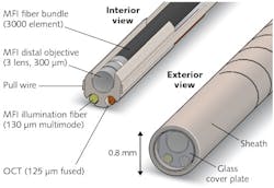
This latest version is a highly flexible, steerable device measuring 0.8 mm in diameter that, unlike MRI, CT, or ultrasound, provides sufficient resolution to detect subtle early changes and can, unlike blood tests, localize the disease (see figure). Using inexpensive optical components, it combines multispectral fluorescence imaging (MFI) and OCT, and works in concert with contrast agents for molecular imaging to generate high-quality images "exquisitely sensitive to early neoplastic changes." The technology has been shown in multiple diseases to have high sensitivity and specificity. The relatively simple technique could be performed under local anesthesia and sedation: an everting balloon will gently open the fallopian tube and guide the falloposcope down the center of the lumen, decreasing the chances of perforation or other injuries. This simplified delivery method could enable use in even small practices, promising broad access to early diagnosis and treatment decision options.

Barbara Gefvert | Editor-in-Chief, BioOptics World (2008-2020)
Barbara G. Gefvert has been a science and technology editor and writer since 1987, and served as editor in chief on multiple publications, including Sensors magazine for nearly a decade.