Imaging Spectrometers: Handheld imaging spectrometer locates nuclear radiation
H3D (Ann Arbor, MI), a spinout from the University of Michigan (also in Ann Arbor), has commercialized a lightweight (9 lb) handheld radiation camera called Polaris-H, a compact imaging spectrometer that can not only identify the isotopic composition of a gamma-ray source, but also image the distributed location of that source.
High-resolution spectrometers identify radiation sources through the detection of high-energy gamma rays. Typical high-resolution spectrometers-using high-purity germanium-can achieve < 0.4% full-width at half-maximum (FWHM) energy resolution at 662 keV, but require cryogenic cooling and cannot perform radiation imaging.
Previously, gamma-ray imaging has been performed in one of three ways. In coded-aperture imaging, a high-atomic-number mask is placed in front of a detector plane, and the shadow cast by the mask on the detector is used to deconvolve the source distribution in the forward direction. However, about half of gamma rays are lost in the mask, reducing imaging efficiency. Imaging can also be performed with a thick collimator on the detector that is rotated through directions of interest; however, detection efficiency is severely degraded, greatly increasing required observation time. Finally, imaging can be performed with two detector planes by recording Compton-scattered pairs between the planes. Since the probability of events with one interaction in each plane is small, the imaging efficiency in this case is also small.
In contrast, the H3D camera is able to image with a single block of crystalline material and no shield. It uses a pixelated cadmium-zinc-tellurium (CdZnTe or "CZT") crystal that achieves nearly the same energy resolution as germanium-based spectrometers for gamma-ray interactions from 50 keV to 3 MeV, but functions as an imaging spectrometer to both identify and image the location of the radiation source. And unlike stilbene or crystal scintillation detection techniques, gamma-ray energy is more accurately determined, and an image of the source distribution is created.
3D position sensing
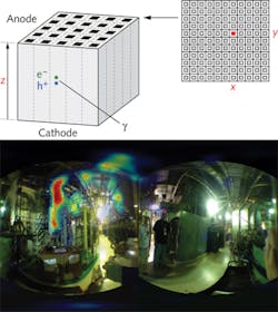
The Polaris-H detector consists of a bulk CZT crystal with a pixelated, patterned anode surface consisting of 121 pixels and bonded to an application-specific integrated circuit (ASIC) chip.
Being highly penetrating, a gamma ray may enter the 20 x 20 x 15 mm CZT crystal from any direction. The gamma ray then undergoes Compton scattering and interacts a second time in the detector. An electric field applied between the anode and cathode sides of the crystal then pulls the resultant charge from each interaction to an anode pixel, identifying its x and y location (see figure). The cathode-to-anode signal ratio or charge drift time is then used to compute the z location or depth of the gamma-ray interaction, producing a 3D map of the interaction energies and positions within the detector. These energies and positions provide information about the direction from which the gamma ray originated.
Data is processed in real time with an embedded computer. The user may view the spectrum and images in real time on a tablet display connected by a cable up to 15 ft from the unit. Transfer through an Internet connection for remote viewing is also available, especially valuable considering that human operators would not want to be in close proximity to strong gamma-ray sources.
To produce an image, the pixel-by-pixel interaction information is processed using a variety of different reconstruction methods. For example, a simple-back-projection (SBP) algorithm uses Compton-imaging techniques that identify the spatial distribution of sources on a mesh surrounding the detector system. The high atomic number of the CZT crystals enables capture of Compton images above several hundred keV.
Although Polaris-H cannot yet reach the < 0.4% FWHM possible with germanium-based spectrometers, current systems come close at 1.1% FWHM at 662 keV for all events and 0.8% FWHM at 662 keV for single-interaction events.
"No one has had a highly portable instrument that they can quickly turn on and use to image particular gamma-ray sources before," says Christopher Wahl, R&D Engineer at H3D. "Now that it is in the field, users are constantly discovering new applications they had never thought possible. Recently, a nuclear power plant discovered a hot spot in a pipe below the floor that they had missed with their other instruments. Now they can shield it and decrease dose to workers."
About the Author

Gail Overton
Senior Editor (2004-2020)
Gail has more than 30 years of engineering, marketing, product management, and editorial experience in the photonics and optical communications industry. Before joining the staff at Laser Focus World in 2004, she held many product management and product marketing roles in the fiber-optics industry, most notably at Hughes (El Segundo, CA), GTE Labs (Waltham, MA), Corning (Corning, NY), Photon Kinetics (Beaverton, OR), and Newport Corporation (Irvine, CA). During her marketing career, Gail published articles in WDM Solutions and Sensors magazine and traveled internationally to conduct product and sales training. Gail received her BS degree in physics, with an emphasis in optics, from San Diego State University in San Diego, CA in May 1986.
